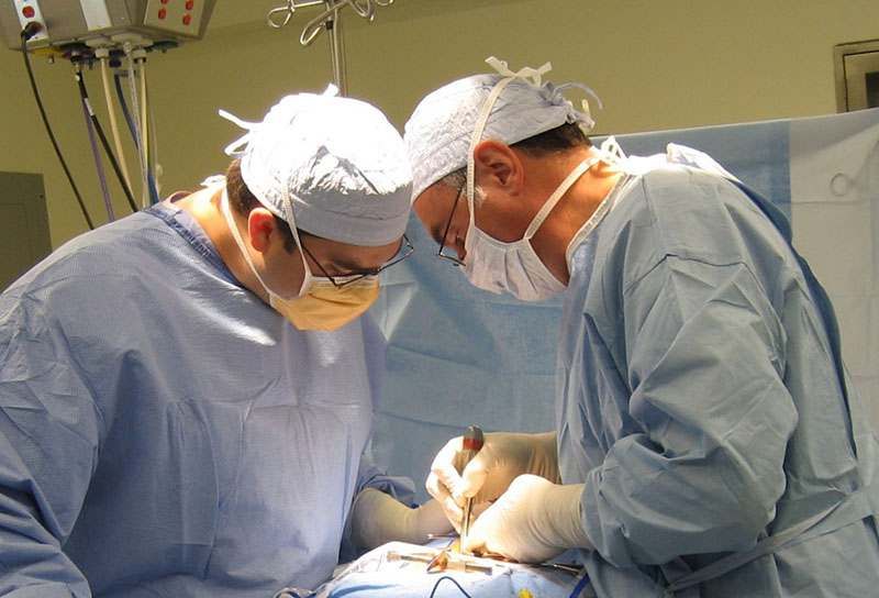
For patients with spondylolisthesis, scoliosis, or degenerative disc disease, spinal fusion is often necessary to correct deformity or stop excessive movement of the vertebrae. Degenerative conditions can cause spinal instability, leading to premature wear and tear of the intervertebral joints and nerve compression. An abnormal curve in the spinal column can cause similar problems.
With spinal fusion, the surgeon ‘locks” two or more vertebrae together in an attempt to increase spinal stability and relieve pain. Fusion does not happen immediately – it is a process that involves the insertion of stabilizing implants and bone grafts to “set” the bone; bony union occurs over a matter of months as the bones mend and grow together.
Procedure overview
There are several ways to achieve spinal fusion, including minimally invasive procedures. Posterior spinal fusion is done after a damaged disc has been removed. Not every fusion requires placement of hardware (i.e., plates, screws), but all require a bone graft. Bone or bone substitutes used in the graft stimulate healing/bone growth, in addition to providing structure/maintaining space between vertebrae.
- The spine is approached from either the front (anterior), back (posterior), or a combination of front/back
- Posterior fusion is usually performed in the upper back (thoracic) and lower back (lumbar) regions.
- Bone for the graft is taken from the patient (autograft) or obtained from a bone bank (allograft), then wedged into place between the vertebrae
- Imaging software and neuro-navigation may be used, depending on the procedure
Treatment of a broken bone in the spine is similar to treatment of a broken bone elsewhere in the body. Spinal fusion immobilizes the vertebrae (much like a cast) so that proper healing can occur.
Posterior spinal fusion typically requires a brief hospital stay, with patients walking the day after surgery. Recovery time is dependent on a variety of factors but ultimately results in regained function and a return to an active quality of life.

