
Microdiscectomy and microdecompression is a minimally-invasive spine surgery that requires a sophisticated surgical microscope, specialized instruments, and microsurgical techniques. Microdiscectomy or microdecompression aims to decompress the nerve root(s) and spinal cord, relieve symptoms, and enable the patient to return to regular activities of daily living. Using advanced minimally-invasive techniques, Dr. Stieber is able to gently spread the muscle surrounding the spine with what is termed sequential tube dilation; this avoids the cutting of muscle generally associated with standard spinal surgery. The use of a robotic microscope allows for excellent visualization of the spine through an extremely small incision, generally 16-18mm.
*This video may potentially be considered graphic to some viewers
Microdiscectomy can be used to treat Herniated Discs.
Possible Benefits of Microdiscectomy and Microdecompression
- tiny incision
- less tissue disruption
- less blood loss
- less post-operative pain
- quicker recovery
Disc degeneration, herniation, and spinal stenosis are common spinal disorders that may compress spinal nerve roots and/or the spinal cord. Compression or pinching of these neurologic structures may result in back and/or neck pain that spreads (radiates) into the lower body (lumbar problems) or upper back, shoulders, and arms (cervical disorders). In addition to pain, neurologic symptoms may develop, including numbness, tingling, and weakness.
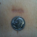
METRx incision 18mm
Not every patient with degenerative disc disease or spinal stenosis requires spine surgery. Dr. Stieber may recommend microdiscectomy or decompression for the following reasons: if non-operative treatment fails to relieve symptoms, progressive worsening of neurologic symptoms or pain, and/or neurological problems. Numbness, loss of function, and weakness are examples of neurologic dysfunction.
About Micro Endoscopic Discectomy and Decompression
Microdiscectomy or microdecompression is performed under general anesthesia. The back is cleansed and prepared for surgery. A tiny skin incision is made approximately the width of a dime. Microsurgical instruments are guided by fluoroscopy (surgical imaging). Sequential dilation with special tubes is performed until the spine can be directly visualized using the operative microscope through a very narrow tube. A small window is made in the bone to access the pinched nerves located within the spinal canal. The nerves are gently moved to the side and protected using specialized microsurgical instruments.
The offending disc fragment or piece of the herniated (ruptured) disc is removed. The nerves are then examined and gently probed to ensure they are free of further compression or problems. The wound is then washed out with antibiotics before the removal of the tube retractor. Once the procedure is complete, the wound is closed with absorbable stitches, and a bandage is placed.
After Surgery
After surgery, the patient is moved into the recovery area. In recovery, nurses and other medical team members closely monitor the patient’s vital signs – pulse, respiration, blood pressure, and pain. Some post-operative incisional discomfort should be expected, and patients receive pain medication either through their IV (intravenous line) or by mouth.
Many patients go home on the same day as surgery. Dr. Stieber discusses the likelihood of hospitalization with patients before their surgery date.
After Care at Home
Dr. Stieber provides microdiscectomy and micro decompression patients with information about what to expect and self-care after hospital discharge. This information includes instructions to control pain, medications, diet, constipation management, bathing and incision care, activity restrictions, and surgical follow-up appointments. As always, Dr. Stieber and his staff welcome questions and ensure that all patient concerns are thoroughly addressed.